Lumps and Bumps by Nancy Kay, DVM
Given the opportunity to examine an older dog, I’ll very likely find at least one or two cutaneous (within the skin) or subcutaneous (just beneath the skin surface) lumps and bumps. Such growths are common by-products of the aging process. In this regard, I liken them to the brown spots that appear on our skin as we get older.
The good news is that most cutaneous and subcutaneous canine tumors are benign. It’s that small population of malignant masses that keeps us on our toes. They are the reason it’s important to have your veterinarian inspect any newly discovered lumps and bumps your dog develops. The smaller a cancerous growth is at the time of treatment, in general, the better the outcome.
Pet your dog!
In terms of “lump and bump patrol,” your first order of business is to pet your dog. No doubt you and your best buddy already enjoy some doggie massage time. What I’m asking you to do is a more methodical petting session. Once a month, slowly and mindfully slide your fingers, palm sides down, along your dog’s body. Move systematically from stem to stern while inspecting for any new lumps or bumps.
Also, look and feel for changes in the size or appearance of those previously discovered. Any new findings should be addressed with your veterinarian who relies upon your help with this surveillance. Imagine your vet trying to find a tiny growth on a shaggy Sheepdog or Sheltie during the course of a single exam. Some lumps and bums are bound to be missed without your assistance.
When to see your veterinarian
Does finding a new growth mean that you must see your veterinarian right away? Not necessarily. Say that you’ve just spotted a new bump in your dog’s skin that is the size of a small pea. She is due for her annual physical examination in three months. Must you go rushing in this week with this new finding, or can it wait the three months? The answer depends on the behavior of this newly discovered growth.
My recommendation is that you continue to observe the new lump once a week. Examining it more frequently can make it difficult to accurately assess change. If the mass is growing, or otherwise changing in appearance, best to have it checked out sooner rather than later. If no changes are observed, waiting to address it at the time of the annual physical exam makes perfectly good sense.
In contrast, say that in the course of examining your best buddy you discover a prune sized, firm, subcutaneous growth that feels attached to her shoulder blade. Based on the larger size and deep attachment of this mass, better to have this one checked out right away. If in doubt, contact your veterinarian to figure out the best course of action. As with most things medical, better to be safe than sorry.
In advance of your veterinary visit, be sure to mark the location of any lumps or bumps requiring inspection. You can clip some hair over the site or mark the fur with a ribbon, hair band, or marking pen. Growths discovered at home when an animal is lying down in a relaxed, comfortable position have a habit of magically disappearing when the dog is upright and uptight in the exam room.
Fine needle aspirate for cytology
If a newly discovered growth is large enough, the usual first step your veterinarian will recommend is a fine needle aspirate for cytology. The purpose of this step is to attempt to noninvasively clarify the cell type within the mass, and whether it is benign or malignant.
Collection of a fine needle aspirate is a simple process that is easy on the dog and rarely requires any sort of sedation. Using a needle no larger than the size of a vaccination needle along with some gentle suction, your vet will remove a smattering of cells from the growth. These cells are then spit out onto a glass slide and evaluated under the microscope.
Some cytology interpretations are a slam-dunk, and can readily be interpreted by your family vet. Others require the eyeballs of a specialist- a clinical pathologist who works in a veterinary diagnostic laboratory. Remember, the goal of the cytology testing is to determine the underlying cell type, therefore whether the growth can be left alone or requires more attention. Fine needle aspirate cytology is often (but not always) definitive. If the results do not provide clarity, a surgical biopsy of the mass may be recommended.
If your veterinarian recommends surgical removal of a mass as the very first step (chooses to forego the fine needle aspirate), I encourage you to consider getting a second opinion. It is always disappointing and frustrating when a veterinarian foregoes cytology, proceeds with surgery, and the biopsy report reveals a malignancy with cancer cells extending beyond the margins of the tissue that was removed. In other words, cancer cells were clearly left behind. Had the veterinarian known in advance from the cytology report that the tumor was malignant, a different approach (much more aggressive surgery and/or radiation therapy) would have been undertaken, almost certainly resulting in a better outcome.
A second “bad news scenario” that can arise from forging ahead with surgery without benefit of fine needle aspirate cytology is failure to identify a cancerous growth that may have already spread elsewhere in the body. If the cytology reveals a malignancy, screening the rest of the body for metastasis (spread) is the logical next step. If metastasis is discovered, removal of the originally discovered mass is unlikely to provide any benefit. Rather, such surgery will only subject the patient (and the client’s pocketbook) to a needless procedure. Leaping into surgery to remove a mass without the benefit of cytology is risky business.
The importance of histopathology
If your veterinarian surgically removes a growth from your dog, do not, I repeat, do not let that tissue sample wind up in the vet clinic garbage can! A far better choice is to have the mass submitted to a veterinary diagnostic laboratory for histopathology (biopsy). There, a veterinary pathologist will evaluate paper-thin slices of the mass under the microscope to confirm the identity of the mass.
Even if a fine needle aspirate cytology indicated that the growth was benign, histopathology is warranted. On occasion, the pathologist discovers something quirky such as a malignant tumor within the center of one that is benign.
If histopathology is not affordable, ask your vet to place the growth that was removed in a small container of formalin (preservative) that you can take home for safekeeping. This way, should multiple masses begin growing at the surgery site or should your dog develop a tumor at another site, you will still be able to request histopathology on the original sample. Formalin is toxic stuff, so keep the container lid sealed tightly.
Lumps and bumps are a very normal part of the canine aging process. Teaming up with your veterinarian to assess them on a regular basis is the very best way to insure that they never create a health issue for your wonderful dog.
Does your dog have any cutaneous or subcutaneous masses? If so, have you had them evaluated by your veterinarian?
If you would like to respond publicly, please visit http://www.speakingforspot.com/blog/?p=4425.
Best wishes for abundant good health,
Dr. Nancy Kay
Diplomate, American College of Veterinary Internal Medicine
Author of Speaking for Spot: Be the Advocate Your Dog Needs to Live a Happy, Healthy, Longer Life
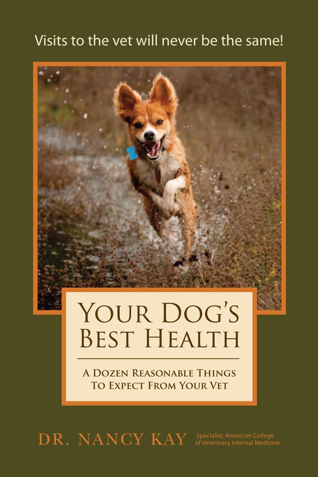
Author of Your Dog's Best Health: A Dozen Reasonable Things to Expect From Your Vet
Recipient, Leo K. Bustad Companion Animal Veterinarian of the Year Award
Recipient, American Animal Hospital Association Animal Welfare and Humane Ethics Award
Recipient, Dog Writers Association of America Award for Best Blog
Recipient, Eukanuba Canine Health Award
Recipient, AKC Club Publication Excellence AwardBecome a Fan of Speaking for Spot on Facebook
Please visit http://www.speakingforspot.com to read excerpts from Speaking for Spot and Your Dog's Best Health. There you will also find "Advocacy Aids"- helpful health forms you can download and use for your own dog, and a collection of published articles on advocating for your pet's health. Speaking for Spot and Your Dog's Best Health are available atwww.speakingforspot.com, Amazon.com, local bookstores, and your favorite online book seller.
|


2 comments:
Excellent book. Excellent information
This was very helpful!
Post a Comment